
Question
The photographs below represent some skeletal materials obtained from a certain mammal.
Study them then answer the questions that follow.
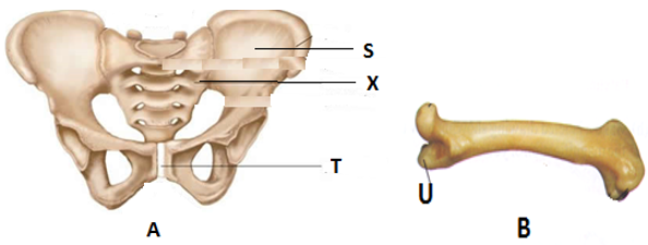
(i) Identify fused bone labelled X
(ii) Name parts S and T on photograph A and part U on photograph B
(iii) Name the type of joint formed at the proximal and distal end of bone B
(iv) Name the type of joint found in structure labelled X
Study them then answer the questions that follow.
(i) Identify fused bone labelled X
(ii) Name parts S and T on photograph A and part U on photograph B
(iii) Name the type of joint formed at the proximal and distal end of bone B
(iv) Name the type of joint found in structure labelled X
Answer
(i) Sacral vertebra
(ii)
S - Illium
T - Pubis symphysis
U - Greater trochanter
Proximal: Ball and socket joint
Distal: Hinge joint