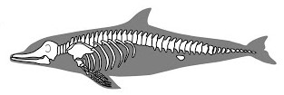
- The mammalian skeleton is divided into two: Axial and appendicular.
-
Axial skeleton is made up of the skull and the vertebral column.
-
Appendicular skeleton is made up of the pelvic and pectoral girdles and limbs (hind limb and forelimbs).
The Axial Skeleton
-This consists of the ;
- Skull
- The Sternum
- Ribs
- The Vertebral Column
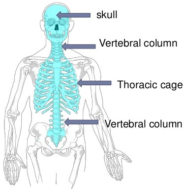
Human Axial Skeleton - Image Courtesy
The Skull
- The skull is made up of cranium and facial bones.
- The cranium; encloses and protects the brain.
- It is made up of many bones joined together by immovable joints.
- The facial bones consists of the upper and lower jaws.
- At the posterior end of the cranium are two smooth rounded protuberances, the occipital condyles.
- These condyles articulate with the atlas vertebra to form a hinge joint, which permits the nodding of the head.
Sternum and ribs
- They form the rib-cage.
- The rib-cage encloses the thoracic cavity protecting delicate organs such as the heart and lungs.
- The ribs articulate with the vertebral column at the back and the sternum at the front.
The Vertebral Column
- The vertebral column is made up of bones called vertebrae placed end to end.
- The vertebrae articulate with one another at the articulating facets.
- In between one vertebra and another is the cartilaginous material called intervertebral disc.
- The discs act as shock absorbers and allow for slight movement.
- Each vertebra consists of a centrum and a neural arch which projects into a neural spine.
- The neural canal is the cavity enclosed by the centrum and the neural arch.
- The spinal cord is located inside the canal.
- The neural spine and other projections e.g. transverse processes serve as points of attachment of muscles.
Type and number of vertebrae in human and rabbit
| Vertebrae |
Human |
Rabbit |
| Cervical |
7 |
7 |
| Thoracic (Neck) |
12 |
12 |
| Lumbar (Upper Abdomen) |
5 |
7 |
| Sacral (Lower Abdomen) |
5 |
3-4 |
| Caudal |
4 (Cocyx) |
16 |
Cervical Vertebrae
- These are found in the neck region of a mammal.
- The distinguishing feature is a pair of vertebrarterial canals in the neural arch, through which the blood vessels of the neck pass.
- Another feature is the structure of the transverse processes.
- They are flattened out and are known as cervical ribs.
- The fIrst cervical vertebra is known as the
Atlas.
- It has a large neural canal and no centrum.
- The second cervical vertebra, is called
axis.
- The other five cervical vertebrae have no specific names.
- They have the same structure.
- The cervical vertebrae possess numerous processes for muscle attachment.
Thoracic Vertebrae
- Each thoracic vertebra has a large centrum ,a large neural canal, neural arch and a long neural spine that projects upwards and backward.
- There is a pair of prezygapophyses and postzygapophyses for articulation with other vertebra .
- They have a pair of short transverse process.
- The thoracic vertebra also articulates with pair of ribs at tubercular and capitular facets.
Lumbar Vertebrae
- Each lumbar vertebra has a large, thick centrum for support of the body.
- It has a neural spine that projects upwards and forwards.
- There is a pair of large transverse process that are directed forwards.
- Above the prezygapophyses lies a pair of processes called metapophyses,
- Below postzygapophyses lies the anapophyses.
- Metapophyses and anapophysis serve for attachment pf muscles of the abdomen.
- In some mammals, there may be another process on lower side of centrum called hypapophysis also for muscle attachment.
Sacral Vertebrae
- The sacral vertebrae are fused together to form a rigid bony structure, the sacrum.
- The centrum of each vertebra is large, but the neural canal is narrow.
- The neural spine is reduced to a small notch.
- The transverse processes of the first sacral vertebra are large and wing-like
- They are firmly attached to the upper part of the pelvic girdle.
Caudal Vertebrae
- Human beings have only four of these vertebrae which are fused together to form coccyx.
- Animals with long tails have many caudal vertebrae.
- A typical caudal vertebra appears as a solid rectangular mass of bone.
- The entire bone consists of the centrum only.
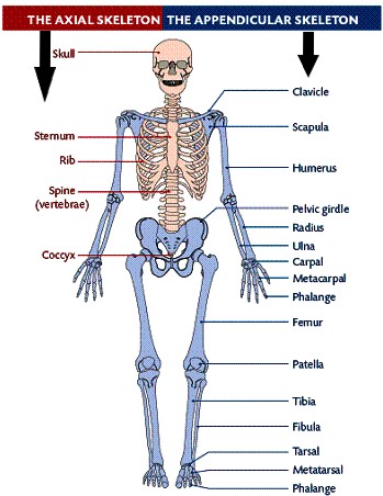
Human Axial and Appendicular Skeleton - Image Courtesy
Appendicular Skeleton
- The appendicular skeleton consist of the limbs and their girdles.
Bones of Fore-limbs
Pectoral girdle
- Pectoral girdle is made of scapula, coracoid and clavicle.
- A cavity known as glenoid cavity occurs at the apex of the scapula.
- The humerus of the fore limb fits into this cavity.
- The clavicle is a curved bone connecting the scapular to the sternum.
Humerus
- Humerus is found in the upper arm.
- It articulates with the scapula at the glenoid cavity of the pectoral girdle and forms a ball and socket joint.
Ulna and radius
- These are two bones found in the forearm.
- The ulna has a projection called olecranon process and a sigmoid notch which articulates with the humerus.
Bones of hind limb
Pelvic Girdle
- The pelvic girdle consists of two halves fused at the pubic symphysis.
- Each half is made up of three fused bones:
- Each half has cup-shaped cavity for the acetabulum for articulation with the head of the femur.
- Between the ischium and pubis is an opening obturator foramen where spinal nerves, blood vessels and a tough inflexible connective tissues pass.
- The ilium, ischium and pubis are fused to form the innominate bone.
The Femur
- The femur is the long bone joining the pelvic girdle and the knee.
- The head of the femur articulates with acetabulum forming the ball and socket joint at the hip.
- The femur has a long shaft.
- At the distal end it has condyles that articulate with the tibia to form a hinge joint at the knee.
- The patella covers the knee joint and prevents the upward movement of the lower leg.
Tibia and Fibula
- The tibia is a large bone, and the fibula a smaller bone is fused to it on the distal part.
- In humans the tibia and fibula are clearly distinguishable.