Esoma-KE Logo
Change Class  |  Change Subject

Plant and Animal Cell - Introduction | The Light Microscope

  Change Class FORM ONE
Select Subject  |  Biology

BIOLOGY REVISION QUESTIONS

Click to Attempt Form 1 Biology Questions


Want to enjoy learning? Try reading without Ads!

Say goodbye to distractions and enjoy uninterrupted learning with our new ad-free feature. Dive into our vast collection of educational content without any interruptions.

Explore Ad-Free learning plus lots of other premium benefits for as little as KES 200 per month.


The Cell: Introduction

- The bodies of living organisms are made up of small microscopic units called cells.
- Some organisms are made up of a single cell only e.g. amoeba and other bacteria in the Kingdom Monera. These organisms are known as unicellular organisms.
- Other organisms are composed of many cells and are said to be multicellular.

- A cell is the basic functional unit of an organism.


Read More On

- Being very small, the cell cannot be seen with a naked eye.
- A powerful magnifying instrument is required.
- The microscope is used to view the cells.

Development of the Light Microscope

- In 1650, Zacharias Jansen invented the compound microscope which combines two lenses for greater magnification.
- In 1665, Robert Hooke used an improved compound microscope to observe cells.

- Between 1650 and 1700, Anthony Van Leewenhoeck developed a better microscope with lenses which provided a greater magnification. He used the microscope to view nuclei and unicellular organisms including bacteria.

- The development of the electron microscope in 1930s significantly improved microbial studies. Through this microscope, it was possible to study very finer details of structures.

Light Microscope

- This is the most commonly used microscope in schools and institutions that do not focus on very fine details of the internal structures of cells.
- The light microscope uses a beam of light to illuminate the specimen being studied.
- A microscope is a delicate and expensive instrument that should be handled with care. It is imperative to understand the parts and functions of various parts of a microscope.

- In a light microscope, the eye piece and the objective lenses both contribute to the magnification of the specimen.

- The total magnification of the specimen viewed under a light microscope will be given by:

Magnification = Eyepiece Lens magnification x Objective Lens Magnification

In particular, if the eyepiece lens magnification is X10 and objective lens magnification power is X8, then the total magnification of the specimen would be:

Magnification = Eyepiece Lens magnification (x10) x Objective Lens Magnification (x8)
= 10 x 8
= X80

Light Microscope - Biology Form One


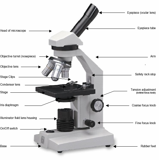
Parts of the Microscope Function of the part
Limb Supports the body tube and stage
Base Provides firm and steady support to the microscope
Body tube Holds the eyepiece and the revolving nosepiece
Coarse adjustment knob Raises or lowers the body tube for longer distance to bring the image into sharper focus
Fine adjustment knob Raises or lowers the body tube for smaller distance to bring the image into sharper focus. It is mostly used with high-power objective lens.
Diaphragm An aperture that regulates the amount of light passing through the condenser to illuminate the specimen.
Eye-piece Contains a lens which contributes to the magnification of the specimen under view.
Objective lens Brings image into focus and magnifies it.
Mirror Reflects light through the condenser to the object on the stage.
Revolving nosepiece Holds the objective lenses in place and enables the change from one objective lens to the other.
Condenser Concentrates light on the object on the stage
Stage Flat platform where the specimen on the slide is placed. It has two clips to hold the slide into position.

Rules of Handling the Light Microscope

  • Always use both hands when carrying the microscope. One hand should hold the base to provide support while the other hand holds the limb.
  • Never place the microscope too close to the edge of the working bench or table.
  • Do not touch the mirror or the lenses with your fingers.
  • Dirty lenses should be cleaned using a special soft lens tissue paper or tissue paper moistened with ethanol. The other parts of the microscope may be cleaned using a microscope.
  • Do not wet any part of the microscope.
  • Make sure the low power objective lens clicks into position in line with the eye piece before and after use.
  • After use, always clean and store the microscope in a safe place, free from moisture and dust.


How to Use the Microscope

  • Place the microscope on the bench with the stage facing away from you.
  • Turn the low power objective lens until it clicks into position.
  • Ensure that the diaphragm is fully open.
  • Look through the eye-piece with one eye; meanwhile adjust the mirror under the stage to ensure that maximum light can pass through. The circular area seen is referred to as the field of view.
  • Again look through the eyepiece while adjusting the mirror under the stage to ensure that sufficient light is passing through the specimen.
  • Use the coarse adjustment knob to bring the low power objective lens to the lowest point. Viewing through the eye-piece, turn the coarse adjustment knob gently until the specimen comes into focus.
  • Use the fine adjustment knob to bring the image into sharp focus. Make a drawing of what you observe.
  • For higher magnifications, turn the medium power objective lens into position and adjust the focus using the coarse adjustment knob. For sharper images, use the fine adjustment knob.
  • If finer details are required, turn the high power objective lens into position; now use only the fine adjustment knob to bring the details into sharper focus.
Cell Structures as seen under the Light Microscope

- The structures within the cell are referred to as organelles. Some of the cell organelles that can be observed under the light microscope include the cell wall, cell membrane, cytoplasm, nucleus, vacuole and chloroplasts.
- These cell organelles perform specific functions within the cell.

Cell Structure - Biology Form One

The cell as seen under the Electron Microscope

Plant Cell Structure under Light Microscope - Biology Form One


Hello Esoma-KE Member, please help review these notes.    Why review

Your review has been successfully submitted.
Tell us what you think about the notes.

Submit Comment

Students' and Teachers' Reviews


Here's what our users had to say ...


Guest User
very fantastic

Guest User
Excellent